About
The Center for Neurological Imaging is a research laboratory at Brigham and Women’s Hospital in Boston. We use quantitative neuroimaging techniques to investigate diseases affecting the central nervous system, with particular emphasis on multiple sclerosis and age-related disorders like Alzheimer’s Disease and cerebrovascular diseases.
Members of the CNI collaborate with other departments within Brigham and Women’s Hospital, with other researchers at Harvard Medical School, with local universities such as Harvard, BU and with gifted clinicians, researchers, and engineers throughout the world.Learn more
Our Projects
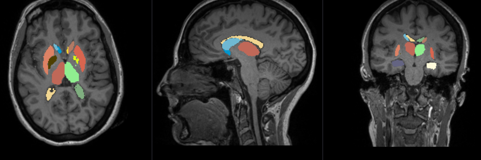
Association of associative, limbic and sensorimotor subcortical structures with fatigue in multiple sclerosis
Context
See also: Project
Discrimination of fatigue and depression networks in patients with multiple sclerosis
Context
See also: Project
Axon-based Parcellation of the Corpus Callosum in Subjects with Multiple Sclerosis
Context
Multiple sclerosis (MS) is a disease affecting approximately 1 out of 1000 people in the U.S. Patients suffer from a variety of neurologic symptoms. The disease typically manifests itself in people in their 20s and 30s, and progresses over years and decades, often severely limiting the independence and productivity…Read more about Axon-based Parcellation of the Corpus Callosum in Subjects with Multiple Sclerosis
See also: Project
Development of a New Method to Measure Glymphatic System Dynamics
Context
Unlike other organs the brain has no lymphatic vessels. In mice, clearance of potentially neurotoxic catabolites is performed through the glymphatic system. Homeostasis between the cerebrospinal fluid (CSF) and the interstitial fluid in the space between the cells of the brain parenchyma drains several metabolites, including beta-amyloid, whose deposition in the cerebral extracellular space has been related to the pathogenesis and progression of Alzheimer’s disease. Very recently, it has been shown that the clearance through the cerebral glymphatic…Read more about Development of a New Method to Measure Glymphatic System Dynamics
See also: Project
Multimodal MRI Approach to Investigate the Development of Brain Damage in Age-related Small Vessel Disease
Context
Cerebral small vessel disease (SVD) is the most common vascular cause of dementia worldwide (Wardlaw et al., 2013), and represents a major determinant of cognitive and physical disability in the aging population (Guttmann et al., 2000; Pantoni, 2010). Several studies, including our own reports (Kaplan et al., 2009; Moscufo et al., 2011, 2012; Papp et al., 2014;…Read more about Multimodal MRI Approach to Investigate the Development of Brain Damage in Age-related Small Vessel Disease
See also: Project
Front- and Back-end Development for SPINE
Context
The “Structured Planning and Implementation of New Explorations” (SPINE) is a web platform designed the to help researches in the design and management the whole lifecycle of a research experiment involving MRI images and clinical data. SPINE aims to answer scientific questions by providing all the required tools to the scientist, enabling experts, such as radiologist, to integrate their knowledge, computer scientist to insert image processing procedures, and statisticians to input models for the analysis of the results, all in the same place. SPINE can…Read more about Front- and Back-end Development for SPINE
See also: Project
Recent Publications
Evaluating the Association between Enlarged Perivascular Spaces and Disease Worsening in Multiple Sclerosis
Michele Cavallari, Svetlana Egorova, Brian C Healy, Miklos Palotai, Juan Carlos Prieto, Mariann Polgar-Turcsanyi, Shahamat Tauhid, Mark Anderson, Bonnie Glanz, Tanuja Chitnis, and Charles RG Guttmann. 2018. “Evaluating the Association between Enlarged Perivascular Spaces and Disease Worsening in Multiple Sclerosis.” J Neuroimaging, 28, 3, Pp. 273-277.Abstract
BACKGROUND AND PURPOSE: Enlarged perivascular spaces (EPVSs) have been associated with relapses and brain atrophy in multiple sclerosis (MS). We investigated the association of EPVS with clinical and MRI features of disease worsening in a well-characterized cohort of relapsing-remitting MS patients prospectively followed for up to 10 years. METHODS: Baseline EPVSs were scored on 1.5T MRI in 30 converters to moderate-severe disability, and 30 nonconverters matched for baseline characteristics. RESULTS: EPVS scores were not significantly different between converters and nonconverters, nor associated with accrual of lesions or brain atrophy. CONCLUSIONS: Our preliminary findings from a relatively small study sample argue against a potential use of EPVS as early indicator of risk for disease worsening in relapsing-remitting MS patients in a clinical setting. Although the small sample size and clinical 1.5T MRI may have limited our ability to detect a significant effect, we provided estimates of the association of EPVS with clinical and MRI indicators of disease worsening in a well-characterized cohort of MS patients.
Dual-Sensitivity Multiple Sclerosis Lesion and CSF Segmentation for Multichannel 3T Brain MRI
Dominik S Meier, Charles RG Guttmann, Subhash Tummala, Nicola Moscufo, Michele Cavallari, Shahamat Tauhid, Rohit Bakshi, and Howard L Weiner. 2018. “Dual-Sensitivity Multiple Sclerosis Lesion and CSF Segmentation for Multichannel 3T Brain MRI.” J Neuroimaging, 28, 1, Pp. 36-47.Abstract
BACKGROUND AND PURPOSE: A pipeline for fully automated segmentation of 3T brain MRI scans in multiple sclerosis (MS) is presented. This 3T morphometry (3TM) pipeline provides indicators of MS disease progression from multichannel datasets with high-resolution 3-dimensional T1-weighted, T2-weighted, and fluid-attenuated inversion-recovery (FLAIR) contrast. 3TM segments white (WM) and gray matter (GM) and cerebrospinal fluid (CSF) to assess atrophy and provides WM lesion (WML) volume. METHODS: To address nonuniform distribution of noise/contrast (eg, posterior fossa in 3D-FLAIR) of 3T magnetic resonance imaging, the method employs dual sensitivity (different sensitivities for lesion detection in predefined regions). We tested this approach by assigning different sensitivities to supratentorial and infratentorial regions, and validated the segmentation for accuracy against manual delineation, and for precision in scan-rescans. RESULTS: Intraclass correlation coefficients of .95, .91, and .86 were observed for WML and CSF segmentation accuracy and brain parenchymal fraction (BPF). Dual sensitivity significantly reduced infratentorial false-positive WMLs, affording increases in global sensitivity without decreasing specificity. Scan-rescan yielded coefficients of variation (COVs) of 8% and .4% for WMLs and BPF and COVs of .8%, 1%, and 2% for GM, WM, and CSF volumes. WML volume difference/precision was .49 ± .72 mL over a range of 0-24 mL. Correlation between BPF and age was r = .62 (P = .0004), and effect size for detecting brain atrophy was Cohen’s d = 1.26 (standardized mean difference vs. healthy controls). CONCLUSIONS: This pipeline produces probability maps for brain lesions and tissue classes, facilitating expert review/correction and may provide high throughput, efficient characterization of MS in large datasets.
Longitudinal microstructural changes of cerebral white matter and their association with mobility performance in older persons
Nicola Moscufo, Dorothy B Wakefield, Dominik S Meier, Michele Cavallari, Charles RG Guttmann, William B White, and Leslie Wolfson. 2018. “Longitudinal microstructural changes of cerebral white matter and their association with mobility performance in older persons.” PLoS One, 13, 3, Pp. e0194051.Abstract
Mobility impairment in older persons is associated with brain white matter hyperintensities (WMH), a common finding in magnetic resonance images and one established imaging biomarker of small vessel disease. The contribution of possible microstructural abnormalities within normal-appearing white matter (NAWM) to mobility, however, remains unclear. We used diffusion tensor imaging (DTI) measures, i.e. fractional anisotropy (FA), mean diffusivity (MD), axial diffusivity (AD), radial diffusivity (RD), to assess microstructural changes within supratentorial NAWM and WMH sub-compartments, and to investigate their association with changes in mobility performance, i.e. Tinetti assessment and the 2.5-meters walk time test. We analyzed baseline (N = 86, age ≥75 years) and 4-year (N = 41) follow-up data. Results from cross-sectional analysis on baseline data showed significant correlation between WMH volume and NAWM-FA (r = -0.33, p = 0.002), NAWM-AD (r = 0.32, p = 0.003) and NAWM-RD (r = 0.39, p = 0.0002). Our longitudinal analysis showed that after 4-years, FA and AD decreased and RD increased within NAWM. In regional tract-based analysis decrease in NAWM-FA and increase in NAWM-RD within the genu of the corpus callosum correlated with slower walk time independent of age, gender and WMH burden. In conclusion, global DTI indices of microstructural integrity indicate that significant changes occur in the supratentorial NAWM over four years. The observed changes likely reflect white matter deterioration resulting from aging as well as accrual of cerebrovascular injury associated with small vessel disease. The observed association between mobility scores and regional measures of NAWM microstructural integrity within the corpus callosum suggests that subtle changes within this structure may contribute to mobility impairment.
Changes to the septo-fornical area might play a role in the pathogenesis of anxiety in multiple sclerosis
Miklos Palotai, Andrea Mike, Michele Cavallari, Erzsebet Strammer, Gergely Orsi, Brian C Healy, Katharina Schregel, Zsolt Illes, and Charles RG Guttmann. 2018. “Changes to the septo-fornical area might play a role in the pathogenesis of anxiety in multiple sclerosis.” Mult Scler, 24, 8, Pp. 1105-1114.Abstract
BACKGROUND: Reports on the relationships between white matter lesion load (WMLL) and fatigue and anxiety in multiple sclerosis (MS) are inconsistent. OBJECTIVE: To investigate the association of total and tract-specific WMLL with fatigue and anxiety. METHODS: Total and regional T2 WMLL was assessed for 19 tracts in 48 MS patients (30 females). ICBM-DTI-81 Atlas-based parcellation was combined with WMLL segmentation of T2-weighted magnetic resonance imaging (MRI). Fatigue, anxiety, and depression were assessed using Fatigue Impact Scale, State Trait Anxiety Inventory, and Beck Depression Inventory, respectively. RESULTS: Fatigue, anxiety, and depression showed significant inter-correlation. We found no association between fatigue and total or regional WMLLs, whereas anxiety was associated with total and regional WMLLs in nine tracts. After adjusting for total WMLL, age, and depression, only the column and body of the fornix (CBF) remained significantly associated with anxiety. Post hoc analyses showed no CBF lesions on T1-weighted MRI and suggested, but could not confirm, that the septum pellucidum might play a role in the pathogenesis of anxiety. CONCLUSION: Our results suggest that anxiety in MS patients may have a neuropathological substrate in the septo-fornical area, which requires further validation using larger sample size and ultra-high-field MRI in targeted prospective studies.
Relationships among clinic, home, and ambulatory blood pressures with small vessel disease of the brain and functional status in older people with hypertension
William B White, Fatima Jalil, Dorothy B Wakefield, Richard F Kaplan, Richard W Bohannon, Charles B Hall, Nicola Moscufo, Douglas Fellows, Charles RG Guttmann, and Leslie Wolfson. 2018. “Relationships among clinic, home, and ambulatory blood pressures with small vessel disease of the brain and functional status in older people with hypertension.” Am Heart J, 205, Pp. 21-30.Abstract
BACKGROUND: Subcortical small vessel disease, represented as white matter hyperintensity (WMH) on magnetic resonance images (MRI) is associated with functional decline in older people with hypertension. We evaluated the relationships of clinic and out-of-office blood pressures (BP) with WMH and functional status in older persons. METHODS: Using cross-sectional data from 199 older study participants enrolled in the INFINITY trial, we analyzed the clinic, 24-hour ambulatory, and home BPs and their relationships with WMH burden and mobility and cognitive outcomes. RESULTS: Volume of WMH was associated with clinic and 24-hour ambulatory systolic BP but not home systolic BP. The mobility measure, supine-to-sit time, had a significant association with 24-hour systolic BP and pulse pressure but not with diastolic BP or values obtained by home BP. Cognitive measures of processing speed (Trails Making Test Part A and the Stroop Word Test) were significantly associated with 24-hour systolic BP, but not clinic and home BPs. CONCLUSION: These data demonstrate that ambulatory BP measurements in older people are more strongly associated with WMH and certain measures of functional status compared to home BP measurements. Hence, home BP may not be a useful substitute for ambulatory BP for assessing subcortical small vessel disease and its consequences. Further longitudinal analyses comparing clinic and various types of out-of-office BP measures with small vessel brain disease are needed. Clinicaltrials.gov identifier: NCT01650402.
Diagnostic value of 3DFLAIR in clinical practice for the detection of infratentorial lesions in multiple sclerosis in regard to dual echo T2 sequences
Salem Hannoun, Damien Heidelberg, Roula Hourani, Thi Thuy Trang Nguyen, Jean-Christophe Brisset, Sylvie Grand, Stéphane Kremer, Fabrice Bonneville, Charles RG Guttmann, Vincent Dousset, and François Cotton. 2018. “Diagnostic value of 3DFLAIR in clinical practice for the detection of infratentorial lesions in multiple sclerosis in regard to dual echo T2 sequences.” Eur J Radiol, 102, Pp. 146-151.Abstract
BACKGROUND AND PURPOSE: The aim of this prospective study is to investigate and evaluate in clinical practice the diagnostic impact of 3DFLAIR in regards to 2DT2/PD in terms of infratentorial lesions detection in multiple sclerosis (MS). MATERIAL AND METHODS: 164 MS patients from the OFSEP database were reviewed retrospectively. MR examinations were performed on 1.5T or 3T systems from four different centers. Infratentorial lesions were counted and allocated to different regions of the posterior fossa by three raters independently (junior resident, resident with an expertise in neuroradiology, and senior neuro-radiologist) on the 3DFLAIR and 2DT2/PD. Both sequences do not have the same spatial resolution but reflect what is recommended by most of the consensus and done in clinical practice. RESULTS: With an overall number of 528 for Rater-1 and 798 for Rater-2 infratentorial lesions, 3DFLAIR had a significantly higher number of lesions detected than 2DT2/PD (303 for Rater-1 and 370 for Rater-2). The prevalence of trigeminal lesions detected by using 3DFLAIR was also significantly higher than 2DT2/PD. ROC analysis showed 3DFLAIR to be more specific and sensitive than 2DT2/PD. An overall difference between all three Raters has been observed. The more the Rater is experienced the more lesions he detects. CONCLUSION: Along with the radiologist ability to detect lesions based on his level of experience, the OFSEP optimized 3DFLAIR can significantly improve infratentorial lesion detection in MS compared to 2DT2/PD. This is important in MS follow-up that takes into account new lesions number to adapt patients’ treatment.
People
Alfredo Morales Pinzon, PhD
Instructor in Radiology, Harvard Medical School
Research Fellow in Radiology, Brigham and Women’s Hospital
Dr. Alfredo Morales Pinzon is an Instructor of Radiology at Brigham and Women’s Hospital and Harvard Medical School working at the Center for Neurological Imaging (CNI). After working in image processing of the lungs using Computed Tomography during his Ph.D., he joint CNI to support and develop image processing pipelines. His work has focused on the development and integration of algorithms with the aim to study the degenerative and reparatory process of tissue brain in the presence of distal white matter lesion using quantitative MRI imaging. His work also includes the development of the Structured Planning and Implementation of New Explorations (SPINE), a virtual laboratory for research…. Read more about Alfredo Morales Pinzon, PhD
Harvard Medical School, Brigham and Women’s Hospital
Department of Radiology,
1249 Boylston Street, Boston, MA, USA
amoralespinzon@bwh.harvard.edu
Yergali Askarbayev
Yergali Askarbayev studied undergraduate in Computing and Software Engineering back in Kazakhstan. After graduation, hw had an experience in an industry as a… Read more about Yergali Askarbayev
Michele Cavallari, MD, PhD
Instructor in Radiology, Brigham and Women’s Hospital
Instructor, Harvard Medical School
Michele Cavallari, MD, PhD is an Instructor in Radiology at the Center for Neurological Imaging. He graduated in Medicine in 2005, then… Read more about Michele Cavallari, MD, PhD
Harvard Medical School, Brigham and Women’s Hospital
Department of Radiology,
1249 Boylston Street, Boston, MA, USA
Charles R. G. Guttmann, MD
Director, Center for Neurological Imaging
Associate Professor of Radiology, Harvard Medical School
Dr. Guttmann is the Director of the Center for Neurological Imaging at Brigham and Women’s Hospital and an Associate Professor of Radiology at Harvard… Read more about Charles R. G. Guttmann, MD
Harvard Medical School, Brigham and Women’s Hospital
Department of Radiology,
1249 Boylston Street, Boston, MA, USA
Andrzej Marciniak, PhD
Postdoctoral Fellow in Radiology, Brigham and Women’s Hospital
Research Fellow, Harvard Medical School
Andrzej Marciniak, PhD, joined the Center for Neurological Imaging team in 2018. His main interest is the development of web-based platform for… Read more about Andrzej Marciniak, PhD
Harvard Medical School, Brigham and Women’s Hospital
Department of Radiology,
1249 Boylston Street, Boston, MA, USA
Nicola Moscufo, PhD
Investigator in Radiology, Brigham & Women’s Hospital
Instructor, Harvard Medical School
Nicola Moscufo, PhD, M.A., joined the Center for Neurological Imaging team in 2004. He is an Investigator in the Department of Radiology,… Read more about Nicola Moscufo, PhD
Harvard Medical School, Brigham and Women’s Hospital
Department of Radiology,
1249 Boylston Street, Boston, MA, USA
Miklos Palotai, MD
Instructor in Radiology, Brigham and Women’s Hospital
Instructor, Harvard Medical School
Miklos Palotai, M.D. is an Instructor in Radiology at the Center for Neurological Imaging (CNI) at Brigham and Women’s Hospital, Harvard Medical… Read more about Miklos Palotai, MD
Harvard Medical School, Brigham and Women’s Hospital
Department of Radiology,
1249 Boylston Street, Boston, MA, USA
Aditya Singh
Research Machine Learning Trainee, Brigham and Women’s Hospital
MS, Boston University
Aditya Singh is pursuing MS with a focus in Machine/Deep Learning working at the Center for Neurological Imaging (CNI). After having worked on…Read more about Aditya Singh
Dana Yespenbetova
Research trainee in Radiology at the Center for Neurological Imaging
Dana Yespenbetova is graduate student at Boston University, majoring in Computer Information Systems. She studied bacherlors of computer science in… Read more about Dana Yespenbetova
Harvard Medical School, Brigham and Women’s Hospital
Department of Radiology,
1249 Boylston Street, Boston, MA, USA
Siddharth Bakshi
Research trainee in Radiology at the Center for Neurological Imaging
Siddharth Bakshi is a graduate student at Boston University, pursuing Data Analytics. He completed his bachelors in Petroleum Engineering from Indian Institute… Read more about Siddharth Bakshi
Giorgio Berardini
Giorgio Berardini is a Master’s degree student in Artificial Intelligence and Machine Learning at University of Calabria, Italy. He is working on automating… Read more about Giorgio Berardini
Avantika Dasgupta
Research trainee in Radiology at the Center for Neurological Imaging
Avantika Dasgupta is a student at Boston University, enrolled in a graduate program in Data Analytics. She earned her bachelors degree in Information… Read more about Avantika Dasgupta
Bissenbay Dauletbayev
Research trainee in Radiology at the Center for Neurological ImagingBissenbay Dauletbayev is a graduate student at Boston…
Read more about Bissenbay Dauletbayev
Harvard Medical School, Brigham and Women’s Hospital
Department of Radiology,
1249 Boylston Street, Boston, MA, USA
Virginia Rinaldi
Research trainee in Radiology at the Center for Neurological Imaging
Virginia Rinaldi is a medical student in her last year of a Medicine Degree Program at Sapienza University of Rome (Sant’Andrea Hospital). She joined the… Read more about Virginia Rinaldi
Harvard Medical School, Brigham and Women’s Hospital
Department of Radiology,
1249 Boylston Street, Boston, MA, USA
Fernanda Troili, MD
Research trainee in Radiology at the Center for Neurological Imaging
Fernanda Troili, MD, is research trainee in Radiology at the Center for Neurological Imaging. She is currently completing her residency in… Read more about Fernanda Troili, MD
Harvard Medical School, Brigham and Women’s Hospital
Department of Radiology,
1249 Boylston Street, Boston, MA, USA
Max Wallack
Research trainee in Radiology at the Center for Neurological Imaging
Max Wallack is a student at Harvard Medical School, class of 2020. He graduated summa cum laude from Boston University in 2015. He previously… Read more about Max Wallack
CONTACT
Dr. Charles R. G. Guttmann, MD
Center for Neurological Imaging
Brigham and Women’s Hospital
1249 Boylston Street
Boston, MA, 02215, USADirections